Bronchoscopy
Bronchoscopy is a diagnostic and therapeutic procedure that allows doctors to directly visualize the inside of the airways (trachea and bronchi) using a thin, flexible tube called a bronchoscope.
Why It's Done (Indications):
- Investigate persistent cough, blood in sputum, or abnormal chest X-ray/CT scan
- Diagnose lung infections, tumors, TB, or interstitial lung disease
- Take biopsies (tissue samples) from the lungs or airways
- Remove foreign bodies, mucus plugs, or tumors
- Lavage (washing out) part of the lung to collect samples
Types of Bronchoscopy:
1. Flexible Bronchoscopy (most common)
- Done under local anesthesia and mild sedation
- Outpatient or short-stay procedure
- Used for diagnostic and minor therapeutic purposes
2. Rigid Bronchoscopy
- Done under general anesthesia
- Used for removing large foreign bodies, tumors, or controlling heavy bleeding
What Can Be Done During Bronchoscopy:
- Bronchoalveolar Lavage (BAL): Saline is sprayed into the lung and collected for testing
- Transbronchial Biopsy: Taking small samples of lung tissue
- Endobronchial Biopsy: Biopsy of visible airway lesions
- Brushings & Washings: Collecting cells for cytology or microbiology
- Foreign body removal
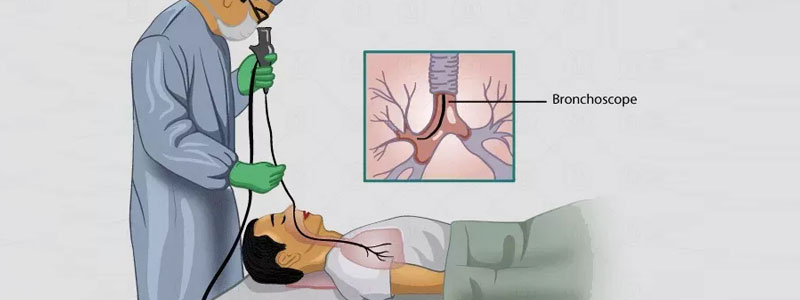
How the Procedure Works:
- Patient is made to lie down or sit upright.
- Local anesthesia is sprayed into the nose or throat.
- A bronchoscope is gently inserted through the nose or mouth, into the windpipe and airways.
- Camera visuals guide the doctor to assess or intervene.
- The procedure usually takes 15–45 minutes.
Aftercare:
- Avoid eating/drinking until the numbing effect wears off (usually a few hours)
- Mild throat discomfort is normal
- Report difficulty breathing, chest pain, or fever immediately